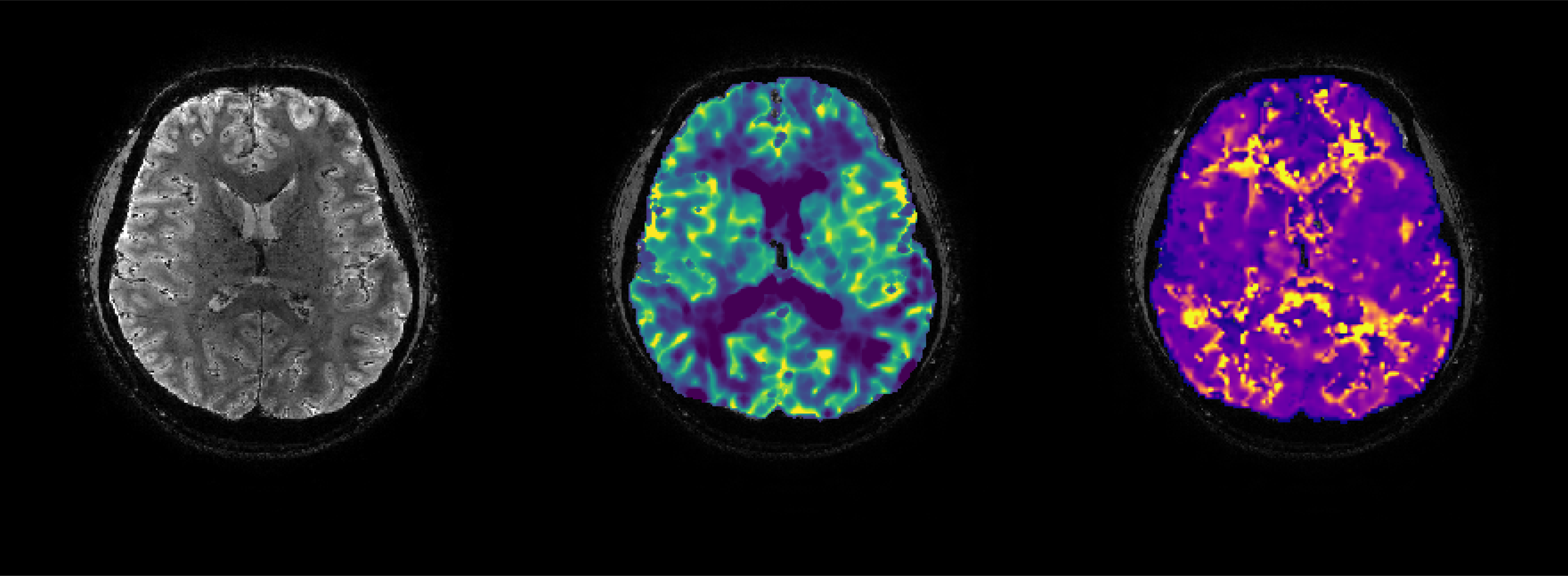
Measuring cerebral blood flow and cerebrovascular reactivity is vital in the pre-operative assessment of moyamoya patients. Over the past two years our team at the UMC Utrecht has been developping a comprehensive multi-modal MRI protocol that includes multi-PLD ASL and BOLD imaging with acetazolamide as an alternative for the commonly used PET-CT method. This approach has spared children harmful radiation and greatly reduced wait times, and has also provided cost savings. Some of these children must be scanned under anaesthesia, which as PhD Candidate Pieter Deckers has discovered can lead to some important caveats for interpreting the clinical MRI scans. Pieter recently presented his work during the Imaging Cerebral Physiology Network (ICP network – not to be confused with insane clown posse 😉 ) webinar. See below!
The seeVR toolbox is being used in an increasing number of studies. I also had the pleasure of giving a presentation for the ICP network where I showed some of the results possible using seeVR ranging from high-resolution cortical measurements to linking the shape of the white matter CVR response with structural properties of the medullary veins. I also presented some exciting work done with the group of Prof. David Mikulis where we used hypoxia to generate arterial BOLD contrast. Here we could implement a carpet-plot analysis (also available in seeVR) to derive measures of blood arrival throughout the brain. Links to the Neuroimage papers and the ICP webinar below!

