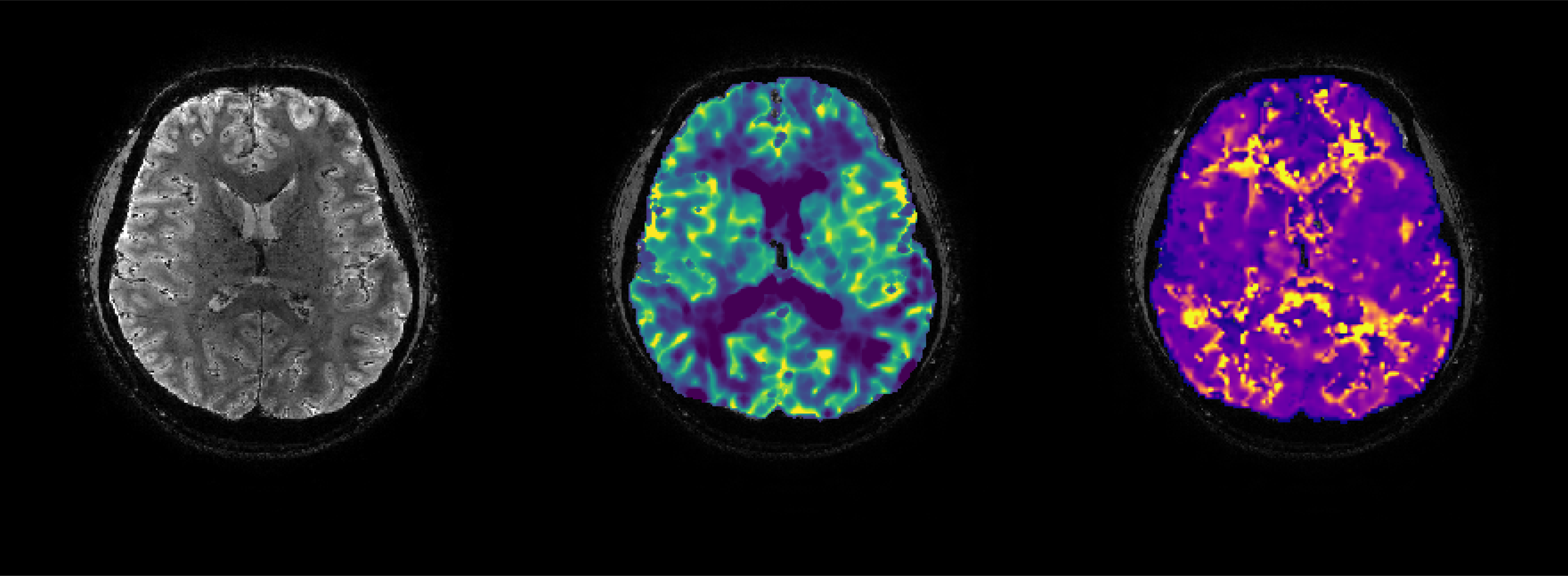
Imaging techniques that are sensitive to hemodynamic status are of high clinical interest for a range of diseases due to their potential for interrogating the presence of collateral blood supply. For instance, collateralization of leptomeningeal vessels is strong predictor of favorable outcome after stroke due to the associated ability to maintain perfusion in the ischemic penumbra for longer periods of time. Similarly, an intact circle-of-Willis and/or strong leptomeningeal collaterals can protect against perfusion deficit in cases of cerebrovascular stenosis. Cerebral auto-regulatory mechanisms play an essential role for capitalizing on alternate blood delivery pathways via vasodilatory responses that serve to draw blood towards affected tissues.
This concept is demonstrated by amazing post-mortem work of Prof. Bart van der Zwan that highlighs the presence of leptomeningeal anastamoses – pial arteries that serve as connecting branches between two major cerebral arteries feeding two different cortical territories. In the video presented below, Dr. van der Zwan injected resin colored with different pigments into the anterior, middle and posterior cerebral arteries. By modulating the input pressure at the large cerebral vessels, it can be seen that blood is able to flow in two directions (green dye passing into the red perfusion territory). In-vivo, this can a provide a protective mechanism driven by the hemodynamic and metabolic needs of the connected territories.
For a nice overview of leptomeningeal collateral function, download the work of Brozici et al. to the right —–>
References
1. van Osch MJ et al., JCBFM 2018, 38(9):1461-1480
2. McVerry F et al., AJNR 2012, 33(3):576-582
3. Van der Zwan et al., Anat Rec. 1990(228):230-236
4. Brozici M et al., Stroke, 2003(34):2750-2762

